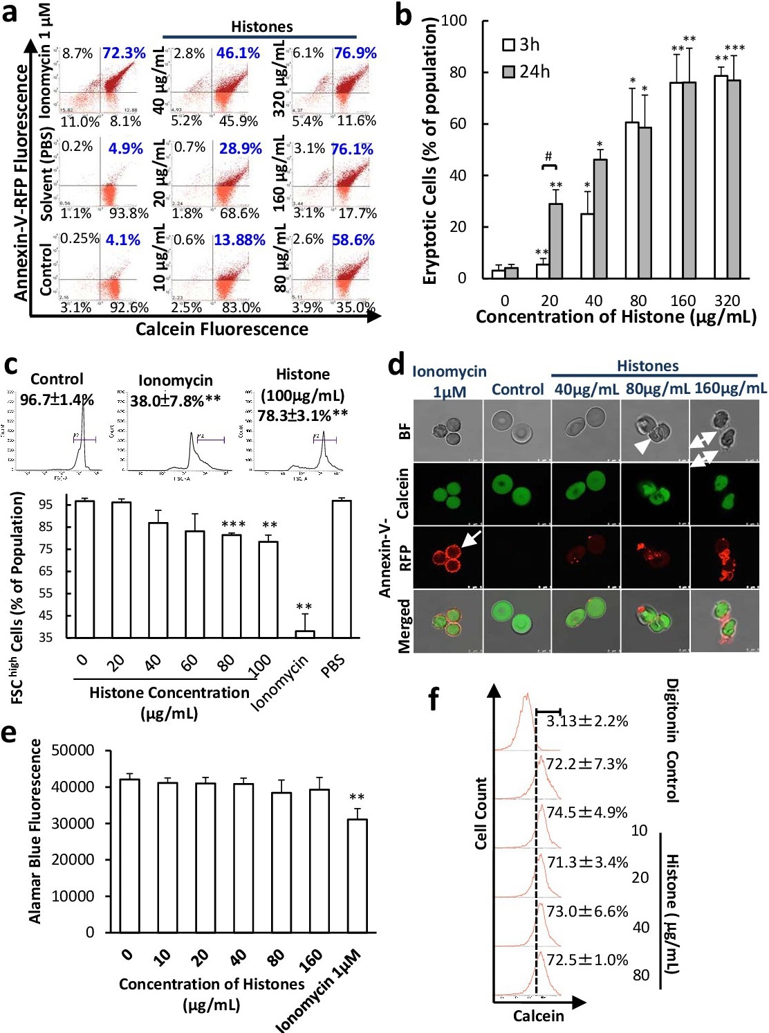
Fig. 1. EHs induce eryptosis in human RBCs. Freshly isolated human RBCs (1.5x10⁷/mL) were treated with the agents as indicated at 37°C for 24 h in a Ca2+-HEPES buffer. After incubation, cells (1x10⁶/mL) were stained with annexin-V-RFP and calcein/AM (1 μM) for 30 min and subjected to flow cytometric or confocal microscopic analysis. (a) Number at the corner representing the % of total population in the corresponding quadrant of the dot plots. (b) Percentage of the early eryptotic cells cells (annexin-Vhigh/calceinhigh) in the flow cytometric analysis after treatment with EHs for 24 (grey bar) or 3 h (white bar). (c) 3-h treatment data for FSC. (d) Confocal microscopic images from cells after 3-h treatment. Echinocytes (star-shape): arrow; stomatocytes (cup-shape): arrow head and membrane blebs: double-arrow. Scale bar: 5 μm. (e) Metabolic activities from cells treated with the agents as indicated at 37°C for 24 h with Alamar Blue. (f) Ghosts entrapped with calcein (non-AM form) were treated with EHs or digitonin (1 μM) at 37°C overnight. Calcein fluorescence was determined by flow cytometry for the degree of membrane permeabilization. Results are mean ± SD (n=3). *p<0.05, **p<0.01, ***p<0.001 relative to the control. #p<0.05, relative to the response of the same concentration with 24-h treatment.